Habit, Habitat and Morphology of the Dogfish

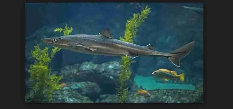
Dogfish are entirely marine and are plentiful in the waters of the European continental shelf. Similar species are found in most parts of the world. They justify their common name by hunting in packs and by their great reliance on their highly – developing sense of smell.
The fishes normally swim near the sea – floor hunting for the crustaceans, molluscs and worms which form the major part of their diet. There are separate sexes, the males and females being easily distinguishable by external as well as internal characteristics. Mating takes place near the surface and each egg is passed out of the body encased in a horny protective covering of distinctive shape. Egg laying takes place throughout the year, except in autumn: the female passing out ten or twelve egg – cases per month. There is no larval stage: the embryo develops within its horny case and the young fish emerges in a well advanced state.
External Features of a Dogfish
The body is about 60cm long, tapering considerably towards the tail. In front, the head is blunt, flattened dorso-ventrally. The trunk region is also flattened ventrally but arched dorsally. Much of the tail is almost cylindrical, the posterior region being somewhat flattened laterally. On the dorsal surface, the body is dark in color, with plentiful brown to black spots. The color fades and the spots diminish down the flanks and the ventral surface is almost white. Covering the body are small sharp spines which all point backward. Breaking up the smooth outline are the fins, of two types. There are four median fins, an anterior dorsal, posterior dorsal, caudal and ventral and two sets of paired fins, pectoral and pelvic.
The head extends from the snout to a point just behind the last gill slit. It is rounded in front and thickens both dorsally and laterally towards its posterior region. The paired eyes are narrow and are placed laterally: they are protected by upper and lower lids, the latter being movable. The wide crescentic mouth is ventral and some distance behind the snout. From it the oronasal grooves pass forward to the rounded nostrils. Behind the eyes are two small round openings called spiracles, which are vestigial gill clefts. Posterior to the spiracles, on each side, is a row of five gill slits, which open from the pharynx to the exterior and are concerned with breathing. Above the gills slits, from a point just behind the eye, a faint lateral line passes down the whole length of the body. It contains sense organs concerned with detection of vibrations in the water: branches of the main lateral line ramify over the head. Scattered in this region are rows of very small pores which lead into ampullae containing mucus. At the bases of these canals are sense organs concerned with perception of temperature changes. There is no neck, the trunk and head being confluence.
The trunk extends from the front of the pectoral fin to the cloaca, where it merges with the tail region. Dorsally and laterally, it is thick and firm, owing to the presence of the myotomes. Ventrally, the muscle is thinner and the body wall is more yielding to the touch. Both pectoral and pelvic fins are attached to the trunk, and between the left and right members of each pair respectively, lies a firm girdle of cartilage. These paired fins are roughly triangular in shape and lie in the horizontal plane. Between the pelvic fins is a shallow depression called the cloaca into which the anus, the reproductive ducts and the urinary papilla open. At the sides of the cloaca are the two small abdominal pores which lead from the splanchnocoel. In the male, two stout curved rods extend backward, one attached to the inner border of each pelvic fin. These are the claspers, which play a part in the mating process: the spines on these point forward.
The tail, which is more than half the length of the body, tapers continuously and is slightly flattened laterally. Apart from the axial skeleton, it is a firm solid mass of muscle and contains none of the viscera. At its posterior end, it is slightly upturned. The four median fins are borne on the tail, the anterior dorsal fin is just beyond the cloacal region, the posterior dorsal is half way between this and the end of the body, while the extensive caudal fin passes along the dorsal surface around the posterior end and then along the ventral surface. This caudal fin is asymmetrical, bearing an extra lobe on the ventral side. Such a tail is said to be heterocercal compared with the symmetrical homocercal tail of many bony fishes. Since the dogfish is heavier than sea – water and has no swim – bladder with which to adjust its relative density, this fin with the extra ventral lobe gives it lift in the water when required. The ventral fin lies in a position mid way between the dorsal fins but on the lower surface. The tail is postanal and metamerically segmented: in the fishes it reaches its greatest development as an organ of locomotion.
Structure and Functions of the Body Wall
In all the vertebrates, the body wall has the same general arrangement of skin, muscle, and the peritoneum abutting on the coelom. The classes show variation mainly in two particulars, the specialized structures develop in the skin and the arrangement of the myotomes. The body wall of the dogfish is very thick and firm dorsally, gradually becoming thinner and less firm towards the soft ventral region.
The skin consists of a stratified epidermis and a thicker mesodermal dermis. This condition contrasts sharply with the invertebrates condition of an epidermis one cell thick and a narrow zone of dermis. The generalized condition of stratified epithelium as described. In the dogfish, the outer cells are dead, flattened and horny: they are continually lost from the surface and replaced by the activity of the stratum germinativum. Small simple saccular mucus glands are plentiful: their secretion affords some measure of protection against infection.
The dermis consists mainly of connective tissues in which bundles of white fibres predominate. During dissection, it will be noticed that the skin is very firmly attached to the underlying muscle. Blood – vessels ramify among the connective tissue and many fine nerves pass through it from the superficial sense organs. Immediately beneath the stratum germinativum are the branched chromatophores containing the pigment melanin. The concentration of these cells determines the colour of the skin. Embedded deeply in the dermis are the dermal denticles or placoid scales, with their points projecting into the water through the epidermis. Each denticle consists of a four – lobbed basal plate connected by a narrower neck to the crown. The basal plate is composed of loose calcified trabeculae, somewhat similar in structure to bone but lacking Haversian systems. The main part of the crown consists of spongy dentine perforated by numerous fine canals in which are protoplasmic fibrils from the cells in the central pulp cavity. The dentine is covered with a thin layer of hard enamel, consisting of hydroxyl – apatite, which infiltrates into the surface of the dentine. In the pulp cavity are the dentine – forming cells called odontoblasts, a small artery an vein with an intervening network of capillaries, a nerve fibr which sends fine branches into the dentine, and delicate areolar connective tissue.
The whole surface is covered with the denticles, which have their points directed backward except on the claspers. There are three distinct types: ventrally they have a single sharp point, dorsally they have three points, while the teeth on the jaws have five points. All are continually replaced, the teeth by the development of new denticles from grooves within the jaws. Comparison with teeth of higher differ from dermal denticles in being attached to the skeleton of the jaws, and in the separate secretion of enamel by the ectoderm.
Beneath the skin lies the muscle of the body wall arranged in myotomes. Viewed from the side with the skin removed, each has a zig – zag shape. Seen in a transverse section, there are a number of separate blocks, each consisting of several concentric rings. It is thus evident that each backward or forward projection of a single myotome is almost a cone, and several closely fitting cones will be cut in one section. The myotomes are connected by myocommata of very tough white fibrous tissue and those of the left and right are joined by a similar vertical sheet. In the trunk region, the innermost connective tissue is joined to the peritoneum, consisting of squamous epithelium which abuts on the coelom.
Functions of the Body Wall of a Dogfish
The skin has protective and sensory functions. Protection is afforded by the horny stratum corneum, by the mucus and by the dermal denticles. The chromatophores produce the resultant dappled color pattern which provides some degree of camouflages. The sensory function is located in the various types of sense organs and nerve endings.
The myotomes, in conjunction with the strong connective tissue sheets and the axial vertebral column, provide for locomotion. The vertebral column is developed in the median sheet of connective tissues: it provides axial firmness together with a limited degree of flexibility.
The peritoneum is a serious membrane which allows almost frictionless sliding of the viscera. In addition, it secretes the coelomic fluid.
Fins are outgrowths of skin into which skeletal tissues penetrates.
Skeleton and Supporting Structures of a Dogfish
The term “skeleton” as used in connexion with vertebrates, usually denotes the bulky endoskeleton. It must be remembered however that there is always some form of exoskeleton, and that sheets of connective tissues and coelomic fluid have a skeletal function. The exoskeleton of the dogfish consists of the stratum corneum and the dermal denticles. The endoskeleton consists of the head which is usually held to include the whole region from the snout to the last gill arch. It consists of the cranium of (brain – case) to which are fused the sense capsules, and the visceral skeleton, a series of arches supporting the tissues between the visceral clefts.
The cranium of the dogfish is a roughly rectangular box which contains the brain. To the front of it are fused the olfactory capsules which h house the olfactory organs, and behind are the auditory capsules which contain the internal ears. Laterally, between the olfactory and auditory capsules are two large depressions called the orbits, bounded dorsally by the supraorbital ridge and ventrally by the infraorbital ridge. The optic capsule is represented by the sclerotic coat of the eye which fits into the orbit. Dorsally, the roof of the cranium is incomplete, being pierced by a large anterior fontanelle in the mid – line behind the olfactory capsules, and by two small foramina between the two auditory capsules, through these foramina the endolymphatic ducts pass to open on the top of the head. In the middle of the supraorbital ridge is a small foramen on each side: through these pass the ophthalmic branches of the fifth and seventh cranial nerves. A foramen is an aperture in the cartilage (or bone) through which a nerve or a blood – vessel passes.
Ventrally, the floor of the cranium is complete: it also forms the roof of the mouth or primary palate. The olfactory capsules have wide ventral openings by which they communicate with the nostrils. On the posterior ventral region are two grooves forming a flattened V with its apex directed forward. In these grooves are the internal carotid arteries which fuse at the apex and pass into the brain through a small foramen. At the outer edges of the grooves are two small foramina through which orbital arteries pass into the orbital sinus surrounding the eye.
Posteriorly, the large foramen magnum is the aperture through which the brain and spinal cord are continuous. At its sides are two round knobs, the occipital condyles, which make a fixed joint with the first vertebra. Lateral to each condyle is a foramen for the teeth cranial nerve, and further to the side, one for the ninth cranial nerve. A small rounded peg in the cartilage between the occipital condyles indicates where the notochord passes into the cranium for a short distance.
A number of foramina in the dogfish are visible in a lateral view of the orbit. The great majority are utilized for cranial nerves. Through the orbito – nasal foramen in the front of the orbit, the olfactory blood sinus is in communication with the orbital sinus. The interorbital canal joins the right and left orbital sinuses, which are also in communication with the anterior cardinal sinus along the post – orbital groove. On the lower side of each auditory capsule is a depression into which the hyomandibular cartilage fits. By this joint the jaws articulate with the cranium.
The Visceral Skeleton of a Dogfish
The visceral skeleton of the dogfish consists of a series of skeletal arches, namely, the mandibular, hyoid and five branchial arches. The mandibular arch on each side consists of two stout bars, an upper palate – pterygo – quadrate and a lower Meckel’s cartilage, constituting the skeleton of the upper and lower jaws. The right and left halves meet in the mid – line in frint.
Each palate – pterygo – quadrate is bound to the cranium by two ligaments, an ethmopalatine near the front, and a spiracular near the rear, it is also bound to part of the hyoid arch, the hyomandibula at the posterior end. Each Meckel’s cartilage articulates with the palate – pterygo – quadrate by a movable joint, and is also bound by a ligament to the hyomandibula. Opening and closing of the mouth is effected only by movement of Meckel’s cartilages. The paired labial cartilages support the corners of the mouth.
The hyoid arch consists of the hyomandibula, a broad ceratohyal which passes within Meckel’s cartilage and amedian flat plate, the basihyal, which supports the tongue. The hyomandibula is the only skeletal attachement of the jaws to the cranium, such a method of jaw suspension being termed hyostylic, in contrast with the autostylic method where the jaws themselves articulate with the cranium.
The five branchiali arches are essentially similar though differeing in details, the most typical or complete being the second. It consist of nine jointed cartilages, movable by muscles. Two pharyngobranchials of the dogfish lie in the roof of the pharynx with their points directed forward, then two epibranchials pass laterally outward to articulate with two ceratobranchials which pass round the pharynx obliquely backward. They articulate ventrally with two short hypobranchials which pass forward to join a single plate, the median basibranchial.


